
Lots of uremic patients use the vascular access to get hemodialysis treatment. The access is like the lifeline of the uremic patient. Hemodialysis patients have dialysis three times a week and must accept cannulations at least six times a week. So the proper cannulation technique is essential as failed punctures have the risk of hematoma and deformation of the access. The use of diagnostic ultrasound to assist needling can reduce puncture failure and help find cannulation sites, effectively improving cannulation quality.
The use of ultrasound-guided cannulation at Hui Der Clinic (輝德診所) has the following advantages:
1. Ultrasounds can accurately evaluate the vascular access. The mature access must comply with Rule of 6s. (1) the depth should not be greater than 0.6cm,(2)the diameter should be at least 0.6cm, (3) access flow should be greater than 600mL/min. The access fistula can only be used six to eight weeks after surgery, and the access graft can only be started three to six weeks after surgery. In the past, physicians used the operation date, auscultation, and the palpation of the access to infer the exact timing of the access cannulation. Now, the use of ultrasound can more accurately evaluate the timing of cannulation.
2. Evaluating the puncture site: Usually, the cannulator can assess the direction of the access by palpation. However, as the access becomes thin or deeper, it is difficult to assess by palpitation. Thankfully, the ultrasound can accurately assess the puncture site. The ultrasound can avoid the risk of bruises or hematoma caused by the cannulation procedure.
How does evaluating a puncture site help?
Example: When a uremic patient who has just transferred from another clinic does not want to be punctured in the same area, we were able to evaluate the puncture site with the help of the ultrasound. In this case, we found an aneurysm. Then, we used an ultrasound to find the most suitable position to perform the buttonhole cannulation technique, as shown in Figure1.
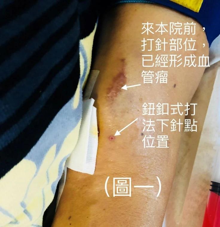
3. Avoid unnecessary repetitive needling: Using an ultrasound, we can evaluate the depth and diameter of the access, allowing us to accurately select the puncture angle and needle size to perform cannulation. Example: When the fistula does not appear on the skin's surface, we used an ultrasound to find the appropriate puncture site, guide the cannulation, and let the needle-tip bypass the blood clot to complete the hemodialysis (shown in Figure II).
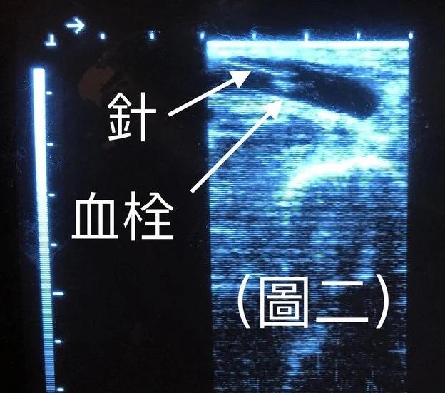
4. Ultrasounds can help adjust the needle during cannulation: The ultrasound-guided needle adjustments reduces the chances of failed punctures for the cannulation.
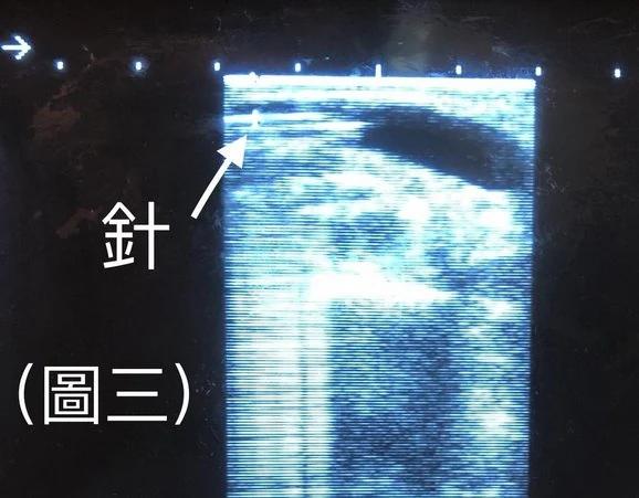
Example 1: A few minutes after the Arterial-Venous fistula set was connected to the tubing set, and the dialysis machine was performed, the arterial side of tubing was found to be flopping around. Everything was going as planned before the tubing was found to be flopping around. Although the patient did not feel uncomfortable, the ultrasound showed (Figure 3), that the needle bevel was upward, and the access was slowly bending into the body. Since the arterial side is pumping blood away from the body, the needle bevel was sucking on the access wall causing the arterial side of tubing to bounce. In such a situation, the physician has chosen to turn the needle over. Fortunately, the problem was solved; there is no longer any bouncing. The venous needle-tip touching the access wall causing pain over the arterial puncture site area was a situation our physicians had to fix using the help of an ultrasound.
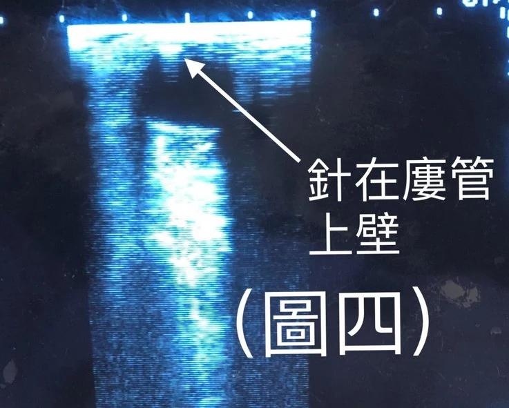
Example 2: In another situation, our patient had a fistula on the left upper-arm. After cannulation, our patient felt pain over the arterial puncture site area; during this time, the dialysis machine is operating normally. The tubing was not bouncing, and the dialysis machine shows that venous pressure is normal. We turned to the ultrasound for help. Upon further inspection, we found that the arterial needle-tip was touching the access wall. We corrected the needle to the proper position in the access by an ultrasound-guided operation. Our patient felt much better; however, the arterial puncture site area was still painful. So, our physician examined the venous needle-tip further. After excluding other possibilities, our physician turned to the ultrasound again. The ultrasound showed the needle-tip touching the upper wall of the access (Figure 4). With the guidance of the ultrasound, we corrected and placed the needle away from the access wall. The patient was immediately relieved from the pain over the arterial puncture side area. In this situation, our patient experienced left shoulder pain. The ultrasound identified the cause of pain as the arterial needle tip touching the access wall of the left upper arm above the elbow. Since patients may not be able to clearly state the reason for the cannulation pain, it is vital to check if the needle-tip is touching the access wall. So for the cases above, ultrasound guidance for adjusting the needle is vital for these conditions.
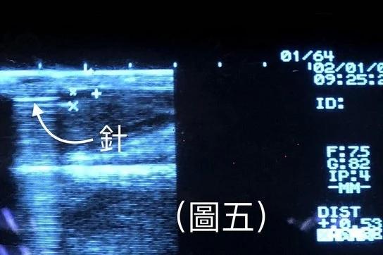
5. Under the guidance of an ultrasound, we can exceed the limits of the Rule of 6, and choose to use the virgin access. Even if we use the virgin access with the standard schedule of cannulation, it is often too deep or to thin to comply with the Rule of 6s. (The graft can be cannulated three to six weeks after the operation, and the fistula can be cannulated six to eight weeks after the operation.) For example (shown in Figure 5), we used ultrasound-guided cannulation to perform hemodialysis on a virgin access that is 0.53cm deep under the skin and about 0.32cm in diameter. Using ultrasound-guided cannulation, we can perform hemodialysis on a virgin access that does not comply with the Rule of 6s. Soon, after using the virgin access, we could also remove the permcath to prevent inflammation, unsmooth blood flow, etc.
6. An ultrasound can identify the problem when there is uncertainty about whether there is a blood clot or obstruction in the access. When we (Hui Der Clinic/輝德診所) use the ultrasound for the access thrombosis, it is usually because of two conditions:
1. The ultrasound confirms that the access is blocked, so there is no need for unnecessary failed cannulations.
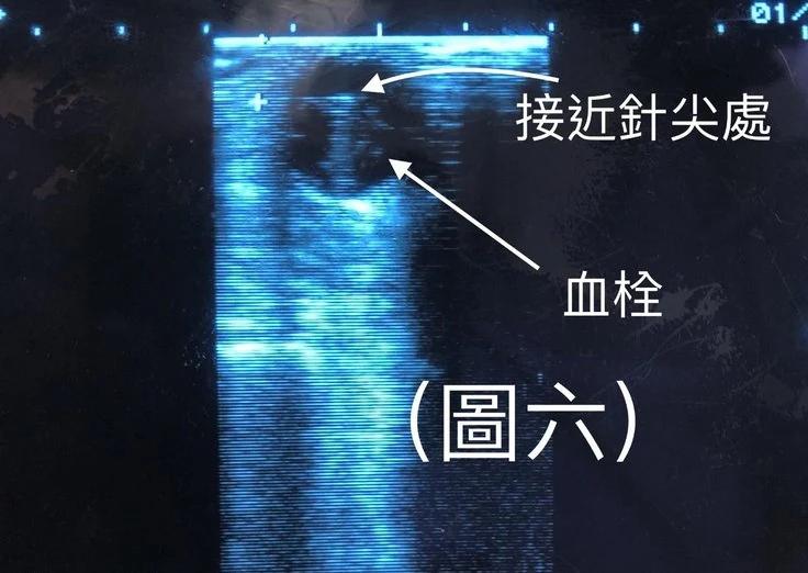
2. When the cannulation is not smooth, using the syringe containing normal saline to test blood reflux cannot draw back blood. There are two additional conditions when the cannulation is not smooth: (1) The ultrasound found that there is a new blood clot of less than 0.5 cm inside the access. We can extract the clot under the guidance of the ultrasound, or we can keep it from this small blood clot to complete the cannulation. (2) The ultrasound found there are many blood clots in the access, and the clots do not completely obstruct the access. Under the guidance of the ultrasound, we can position the needle-tip in the access where there are relatively fewer clots and smoothly complete the hemodialysis, as shown in figure 6. Hui Der Clinic (輝德診所), had the experience of using heparin to treat blood clots in the access during hemodialysis. Blood clots in the access almost completely disappeared after 4 hours of ultrasound tracking.
From our experience, cannulation with or without ultrasound guidance is equally important.
Needling directly (without using ultrasound-guided techniques) can save time; for the easily accessible access, choosing ultrasound-guided cannulation is a waste of time. However, for a difficult to approach access, cannulation without an ultrasound has a higher chance of failure.
Ultrasound-guided cannulation is an excellent technique for the remote access. Though it takes relatively longer to complete, ultrasound guidance can provide the real-time needle position to avoid damaging the access.
Unsuccessful cannulations can cause irritability and fear in patients who are upset to know that they must accept hemodialysis treatment. Using an ultrasound can lower the chances of unsuccessful cannulations, and reduce the pressure patients face during the cannulation. I, Doctor Han Yun Kai, sincerely hope that this technology can be used to benefit patients.